

Volume 23, Issue 4 (October & November 2020)
J Arak Uni Med Sci 2020, 23(4): 512-523 |
Back to browse issues page


![]()
![]()
![]()
Download citation:
BibTeX | RIS | EndNote | Medlars | ProCite | Reference Manager | RefWorks
Send citation to:


BibTeX | RIS | EndNote | Medlars | ProCite | Reference Manager | RefWorks
Send citation to:
Hemmati H, Aboutalebi J, Farzin M, Hemmati G, Rafiei E. Evaluation of Demographic and Clinical Characteristics of the Patients Undergoing Surgery With Ultrasound in Internal Jugular Vein Cannulation Requiring Central Venous Catheter. J Arak Uni Med Sci 2020; 23 (4) :512-523
URL: http://jams.arakmu.ac.ir/article-1-6238-en.html
URL: http://jams.arakmu.ac.ir/article-1-6238-en.html


1- Razi Clinical Research Development Unit, Razi Hospital, Guilan University of Medical Sciences, Rasht, Iran. , drhossein.hemmati@gmail.com
2- Razi Clinical Research Development Unit, Razi Hospital, Guilan University of Medical Sciences, Rasht, Iran.
2- Razi Clinical Research Development Unit, Razi Hospital, Guilan University of Medical Sciences, Rasht, Iran.
Full-Text [PDF 3574 kb]
(1243 Downloads)
| Abstract (HTML) (3791 Views)
Full-Text: (1642 Views)
1. Introduction
nternal jugular vein cannulation is a common procedure for access to the central vein [1]. In most patients, access to the central vein is required [2]. The most common indications for intravenous drugs are high concentrations [3]. The catheter position is between the lower third of the superior vena cava and the upper third of the right atrium [4]. It is recommended that venipuncture be performed under ultrasound guidance [4]. Safe punctures using anatomical landmarks have been introduced since 1966 [5, 6]. Catheters also have complications. Air embolism arrhythmia occurs [7, 8].
In the installation of a central catheter, the ultrasound method is not possible for reasons such as the unavailability of instruments and the lack of trained personnel [9, 10]. The use of ultrasound is not common in Iran [11]. Ultrasound catheterization took less time to reach the jugular vein [12]. Ultrasound increases the success rate and reduces problems [13]. Ultrasound is preferred in infants and children [14]. In this study, demographic and clinical characteristics of patients undergoing ultrasound in internal jugular vein cannulation were examined.
In the installation of a central catheter, the ultrasound method is not possible for reasons such as the unavailability of instruments and the lack of trained personnel [9, 10]. The use of ultrasound is not common in Iran [11]. Ultrasound catheterization took less time to reach the jugular vein [12]. Ultrasound increases the success rate and reduces problems [13]. Ultrasound is preferred in infants and children [14]. In this study, demographic and clinical characteristics of patients undergoing ultrasound in internal jugular vein cannulation were examined.
2. Materials and Methods
In this prospective cross-sectional study, 100 patients referred to Rasht city’s Razi Hospital (2017), who were candidates for central venous catheter, were included in the study by census method [12, 15]. The inclusion criteria consisted of several patients referred for central venous catheter implantation, including patients with local or systemic infection, vascular abnormalities, untreated coagulation disorder (platelets <50000/mm3, and PT or aPTT (or both) disorders. The exclusion criteria included patient death during catheterization.
In this study, 100 patients were randomly selected. Characteristics of the groups (including age, sex, BMI, catheter placement, neck type, the distance between two areas determined by anatomical landmark and ultrasound, and variation of internal jugular vein relative to the carotid artery) were recorded in a researcher-made form and then reviewed and analyzed upon completion of the project. The sampling method was conducted using a convenience sampling. SPSS V. 18 software was used to analyze the results. P<0.05 was considered significant.
In this study, 100 patients were randomly selected. Characteristics of the groups (including age, sex, BMI, catheter placement, neck type, the distance between two areas determined by anatomical landmark and ultrasound, and variation of internal jugular vein relative to the carotid artery) were recorded in a researcher-made form and then reviewed and analyzed upon completion of the project. The sampling method was conducted using a convenience sampling. SPSS V. 18 software was used to analyze the results. P<0.05 was considered significant.
3. Results
In this study, out of 100 patients, only 46 patients (46%) had a normal BMI (Table 1).

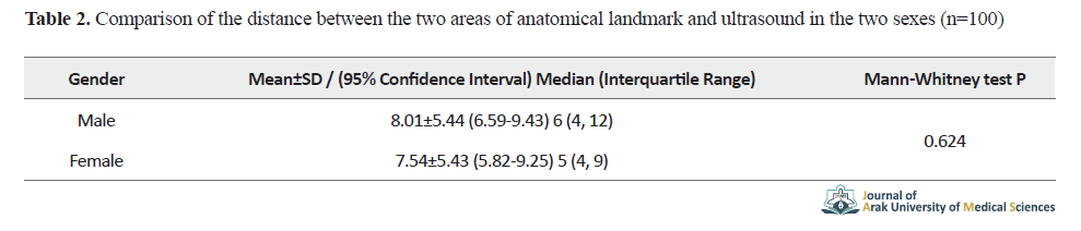
There was no statistically significant correlation between age and the distance between the two areas of anatomical landmark and ultrasound. The distance between the two areas of anatomical landmark and ultrasound was greater in male patients. No statistically significant difference was observed (Table 2).
There was a weak positive correlation between BMI and the distance between the two areas of anatomical landmark and ultrasound, but this correlation was not statistically significant (Table 3). The distance between the anatomical landmark and ultrasound in obese patients was not statistically significant (Table 3).
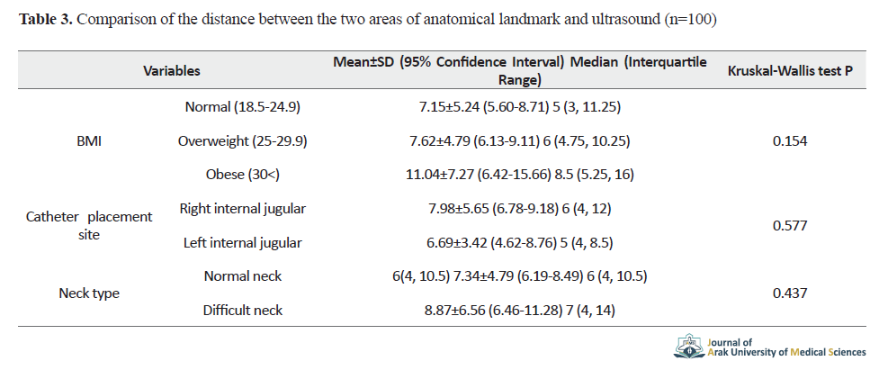
The results showed that the distance between the two areas of anatomical landmark and ultrasound was greater in patients with catheter placement in the right internal jugular vein than in patients with catheter placement in the left internal jugular vein, but this difference was not reported statistically significant (Table 3). The results also showed that the distance between the two areas of anatomical landmark and ultrasound in patients with difficult necks was higher than those with normal necks, but this difference was not statistically significant (Table 3).
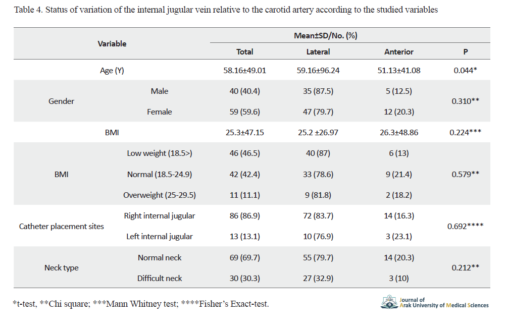
The age of patients in the position of variation of the internal jugular vein relative to the carotid artery was higher than that of the lateral carotid artery compared to the anterior, and in this case a statistically significant difference was observed. According to the results, the variation of the internal jugular vein was not statistically significant in terms of gender, BMI, catheter placement, and neck type (Table 4).
There was no statistically significant correlation between age and the distance between the two areas of anatomical landmark and ultrasound. The distance between the two areas of anatomical landmark and ultrasound was greater in male patients. No statistically significant difference was observed (Table 2).
There was a weak positive correlation between BMI and the distance between the two areas of anatomical landmark and ultrasound, but this correlation was not statistically significant (Table 3). The distance between the anatomical landmark and ultrasound in obese patients was not statistically significant (Table 3).
The results showed that the distance between the two areas of anatomical landmark and ultrasound was greater in patients with catheter placement in the right internal jugular vein than in patients with catheter placement in the left internal jugular vein, but this difference was not reported statistically significant (Table 3). The results also showed that the distance between the two areas of anatomical landmark and ultrasound in patients with difficult necks was higher than those with normal necks, but this difference was not statistically significant (Table 3).
The age of patients in the position of variation of the internal jugular vein relative to the carotid artery was higher than that of the lateral carotid artery compared to the anterior, and in this case a statistically significant difference was observed. According to the results, the variation of the internal jugular vein was not statistically significant in terms of gender, BMI, catheter placement, and neck type (Table 4).
4. Discussion and Conclusion
The present study was performed on 100 patients referred to Razi Hospital in Rasht city, to examining the demographic and clinical characteristics of these patients undergoing ultrasound in internal jugular vein cannulation requiring a central venous catheter. The results regarding the variation of the internal jugular vein in terms of gender, BMI, catheter placement and neck type were not statistically significant. They were not related to the distance between the two markers.
There was no statistically significant difference between the two areas of anatomical landmark and ultrasound in men compared to women. However, there was a statistically significant difference regarding the age of the patients. The use of anatomical landmarks and their results have been reported in several studies.
In the Karakitsos study [16], a successful cannulation rate of 94.4% was reported, which was consistent with the success rate in previous reports [17]. The distance between landmarks in these two methods was statistically different but not significant. In previous studies, only ultrasound guidance was performed [18]. It was performed in patients with severe illness or patients with mechanical ventilation. Our study showed that there was a gap between the anatomical landmark (conventional method) and the ultrasound landmark. Based on previous studies and approval of the reduction of catheterization complications using ultrasound, in the current study, due to the possibility of determining the exact location, catheterization with ultrasound was safer.
In a prospective study by Turker et al. [19], the superiority of cannulation with internal jugular vein ultrasonography was demonstrated. In most studies, central venous catheters have been placed using ultrasound guidance alone. Central venous catheters were performed for 3 minutes for all patients with an ultrasound guide. The results of our study showed that internal jugular vein cannulation with ultrasound had a shorter access time and lower complication rate.
Ethical Considerations
Compliance with ethical guidelines
This research ethically approved by the Ethics Committee of Guilan University of Medical Sciences (Code: IR.GUMS.REC.1396.20).
Funding
This article was extracted from PhD. dissertation of the second author, at the Razi Clinical Research Development Unit, Razi Hospital, Guilan University of Medical Sciences.
Authors' contributions
All authors met the standard writing criteria based on the recommendations of the International Committee of Medical Journal Publishers (ICMJE) and all contributed equally to the writing of the work.
Conflicts of interest
The authors declared no conflict of interest.
Acknowledgements
We would like to thank the managers and colleagues of all departments of the faculties, students, Razi Clinical Research Development Unit, Razi Hospital of the University of Medical Sciences, and those who collaborated in this project.
Send email to the article author
| Rights and permissions | |
 |
This work is licensed under a Creative Commons Attribution-NonCommercial 4.0 International License. |
